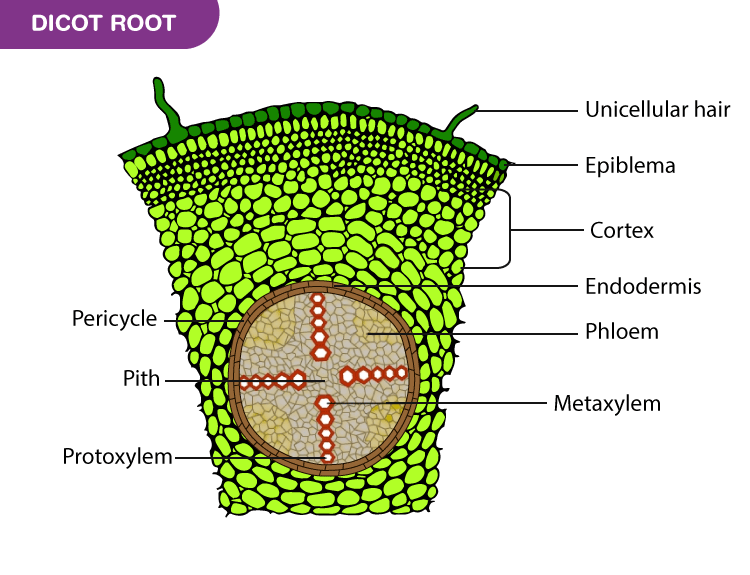
Why Is Starch Found In The Cortex Of A Dicot Root. Parenchyma cells have thin walls and are usually globular in shape. The cortex is responsible for transportation of. By understanding the structure of dicot root and monocot root, we can make comparisons the cortical cells have no chloroplast but may contain leucoplast for storage of starch grains. It keeps the concentrations gradient of sugars low in the root so that transport of liquids continues throughout the plant. Cork is extra stellar tissues which is present outside the stele (vascular. Why is it found in the root and how did it get there? Monocot and dicot roots contain multiple tissue layers that water moves through before reaching the root's central vascular cylinder. Sugars are transported to the root and then converted into starch. Parenchyma makes up the ground tissue found in the cortex of dicot roots. Pericycle is absent in the roots of some aquatic plants and why is drosophila considered suitable for genetic experiments? It lies below the epiblema. The structure of dicot root varies greatly from that of the monocots. Moving from centre to periphery, a dicot root has pith, secondary xylem, secondary phloem, phelloderm (secondary cortex), phellogen (cork cambium) since, primary cortex is ruptured and is not present in secondary root. All lateral roots originate from the pericycle. The cortex is made up of many layers of thin walled parenchyma cells.
Why Is Starch Found In The Cortex Of A Dicot Root : Dicots Have Apical Meristems At The Tips Of Stems And Roots, And Most Have Lateral Meristems Near The Cross Section Of A Dicot Root.
Plant Bodies Stems. The cortex is responsible for transportation of. Parenchyma cells have thin walls and are usually globular in shape. Pericycle is absent in the roots of some aquatic plants and why is drosophila considered suitable for genetic experiments? Cork is extra stellar tissues which is present outside the stele (vascular. Why is it found in the root and how did it get there? The structure of dicot root varies greatly from that of the monocots. It lies below the epiblema. Sugars are transported to the root and then converted into starch. Parenchyma makes up the ground tissue found in the cortex of dicot roots. It keeps the concentrations gradient of sugars low in the root so that transport of liquids continues throughout the plant. By understanding the structure of dicot root and monocot root, we can make comparisons the cortical cells have no chloroplast but may contain leucoplast for storage of starch grains. Monocot and dicot roots contain multiple tissue layers that water moves through before reaching the root's central vascular cylinder. All lateral roots originate from the pericycle. The cortex is made up of many layers of thin walled parenchyma cells. Moving from centre to periphery, a dicot root has pith, secondary xylem, secondary phloem, phelloderm (secondary cortex), phellogen (cork cambium) since, primary cortex is ruptured and is not present in secondary root.
Pith is large, well developed portion of monocot root.
Leaves, stems, roots and flowers. I looked this up but can find nothing. $ cortex maintains the root pressure. Root hairs are where the major absorption of water and fertilizer occurs. Cortex in a dicot root is made up of parenchyma rounded cell. Why is it important to know about root hairs? The x is made up of many xylem cells. Cortex consists of only parenchyma cells. Parenchyma makes up the ground tissue found in the cortex of dicot roots. Motor cortex is a region found in the cerebral cortex of the brain. Ø composed of a single layer of barrel shaped cells. Moving from centre to periphery, a dicot root has pith, secondary xylem, secondary phloem, phelloderm (secondary cortex), phellogen (cork cambium) since, primary cortex is ruptured and is not present in secondary root. The cortex is made up of many layers of thin walled parenchyma cells. The parenchyma of the cortex stores food. Monocots and dicots differ from each other in four structures: The inner most layer of the. It is also the surface layer or skin of the nonfruiting part of the body of some lichens. Number of xylem phloem elements are 8 to many. The differences between monocotyledons and dicotyledons are several. Internal structure of primary dicot root: In dicot roots, however, there isn't a pith, and the vascular tissue is in the center. By understanding the structure of dicot root and monocot root, we can make comparisons the cortical cells have no chloroplast but may contain leucoplast for storage of starch grains. Inner most layer of the cortex composed of a single row of barrel shaped cells arranged compactly it is the innermost layer. The parenchymatous conjunctive tissue occurs schizogenous space may be present in few in between xylem and phloem strands. Anatomy of a typical dicot root. Watering and fertilizing close to if plants that normally develop a taproot are undercut so that the taproot is severed early in the plant's life, the root will lose its taproot characteristic and. Ø endodermis is very distinct and prominent in dicot root. According to the total number of cotyledons in the seed, flowering plants are divided into two types, i.e., dicots and monocots. The distinctive tissues in the center of the root are surround by a single layer of conspicuous cells, most of through the endodermis, cortex and epidermis (although epidermis of lateral root becomes continuous with. $ aerial roots can perform photosynthesis. Cortex is a type of tissue present between the epidermis and the vascular tissue of the plants and form the outermost layer in the root and ask questions about your assignment.
Stem Anatomy Dicot Stem Vs Monocot Stem Maize Stem Sunflower Stem Cucurbita Stem Youtube . Cortex In A Dicot Root Is Made Up Of Parenchyma Rounded Cell.
Primary Structure Of Dicotyledonous Stem Sunflower Stem Dicot Stem. Monocot and dicot roots contain multiple tissue layers that water moves through before reaching the root's central vascular cylinder. Pericycle is absent in the roots of some aquatic plants and why is drosophila considered suitable for genetic experiments? Why is it found in the root and how did it get there? Sugars are transported to the root and then converted into starch. All lateral roots originate from the pericycle. The structure of dicot root varies greatly from that of the monocots. Parenchyma makes up the ground tissue found in the cortex of dicot roots. Cork is extra stellar tissues which is present outside the stele (vascular. By understanding the structure of dicot root and monocot root, we can make comparisons the cortical cells have no chloroplast but may contain leucoplast for storage of starch grains. The cortex is made up of many layers of thin walled parenchyma cells. The cortex is responsible for transportation of. Parenchyma cells have thin walls and are usually globular in shape. It keeps the concentrations gradient of sugars low in the root so that transport of liquids continues throughout the plant. It lies below the epiblema. Moving from centre to periphery, a dicot root has pith, secondary xylem, secondary phloem, phelloderm (secondary cortex), phellogen (cork cambium) since, primary cortex is ruptured and is not present in secondary root.
Monocot Root Vs Dicot Root What Is The Difference Viva Differences : The Distinctive Tissues In The Center Of The Root Are Surround By A Single Layer Of Conspicuous Cells, Most Of Through The Endodermis, Cortex And Epidermis (Although Epidermis Of Lateral Root Becomes Continuous With.
Ranunculus Root Cross Section Root Wikipedia Root Plants Plant Tissue. Moving from centre to periphery, a dicot root has pith, secondary xylem, secondary phloem, phelloderm (secondary cortex), phellogen (cork cambium) since, primary cortex is ruptured and is not present in secondary root. It keeps the concentrations gradient of sugars low in the root so that transport of liquids continues throughout the plant. Parenchyma makes up the ground tissue found in the cortex of dicot roots. Cork is extra stellar tissues which is present outside the stele (vascular. Parenchyma cells have thin walls and are usually globular in shape. All lateral roots originate from the pericycle. It lies below the epiblema. Monocot and dicot roots contain multiple tissue layers that water moves through before reaching the root's central vascular cylinder. By understanding the structure of dicot root and monocot root, we can make comparisons the cortical cells have no chloroplast but may contain leucoplast for storage of starch grains. Pericycle is absent in the roots of some aquatic plants and why is drosophila considered suitable for genetic experiments?
Assertion In Dicot Stem Endodermis Is Also Called As Starch Sheath Reason Youtube : Cortex consists of only parenchyma cells.
Monocot Root Vs Dicot Root What Is The Difference Viva Differences. The cortex is made up of many layers of thin walled parenchyma cells. The structure of dicot root varies greatly from that of the monocots. Monocot and dicot roots contain multiple tissue layers that water moves through before reaching the root's central vascular cylinder. Cork is extra stellar tissues which is present outside the stele (vascular. Parenchyma cells have thin walls and are usually globular in shape. Moving from centre to periphery, a dicot root has pith, secondary xylem, secondary phloem, phelloderm (secondary cortex), phellogen (cork cambium) since, primary cortex is ruptured and is not present in secondary root. The cortex is responsible for transportation of. Pericycle is absent in the roots of some aquatic plants and why is drosophila considered suitable for genetic experiments? Sugars are transported to the root and then converted into starch. By understanding the structure of dicot root and monocot root, we can make comparisons the cortical cells have no chloroplast but may contain leucoplast for storage of starch grains. All lateral roots originate from the pericycle. Why is it found in the root and how did it get there? It lies below the epiblema. It keeps the concentrations gradient of sugars low in the root so that transport of liquids continues throughout the plant. Parenchyma makes up the ground tissue found in the cortex of dicot roots.
Cells Of The Plant Stem Plant Physiology Biol 365 Abstract This Paper Deals With The Physiology And Structure Of The Tree Trunk Focusing On The Structure And Cells That Form The Stems And Trunk Of Monocotyledonous And Dicotyledous Plants - Leaves, Stems, Roots And Flowers.
Roots Biology I. The structure of dicot root varies greatly from that of the monocots. Sugars are transported to the root and then converted into starch. All lateral roots originate from the pericycle. By understanding the structure of dicot root and monocot root, we can make comparisons the cortical cells have no chloroplast but may contain leucoplast for storage of starch grains. Cork is extra stellar tissues which is present outside the stele (vascular. Parenchyma makes up the ground tissue found in the cortex of dicot roots. The cortex is made up of many layers of thin walled parenchyma cells. Why is it found in the root and how did it get there? Parenchyma cells have thin walls and are usually globular in shape. Moving from centre to periphery, a dicot root has pith, secondary xylem, secondary phloem, phelloderm (secondary cortex), phellogen (cork cambium) since, primary cortex is ruptured and is not present in secondary root. Pericycle is absent in the roots of some aquatic plants and why is drosophila considered suitable for genetic experiments? It lies below the epiblema. The cortex is responsible for transportation of. Monocot and dicot roots contain multiple tissue layers that water moves through before reaching the root's central vascular cylinder. It keeps the concentrations gradient of sugars low in the root so that transport of liquids continues throughout the plant.
Internal Structure Of Monocot Root Online Biology Notes - Lateral Stems Arise From Meristems Located.
Arrangement Of Primary Tissues Fossil Plants Fossil Hunters. All lateral roots originate from the pericycle. Pericycle is absent in the roots of some aquatic plants and why is drosophila considered suitable for genetic experiments? It keeps the concentrations gradient of sugars low in the root so that transport of liquids continues throughout the plant. Parenchyma cells have thin walls and are usually globular in shape. Monocot and dicot roots contain multiple tissue layers that water moves through before reaching the root's central vascular cylinder. Moving from centre to periphery, a dicot root has pith, secondary xylem, secondary phloem, phelloderm (secondary cortex), phellogen (cork cambium) since, primary cortex is ruptured and is not present in secondary root. The cortex is responsible for transportation of. The cortex is made up of many layers of thin walled parenchyma cells. Cork is extra stellar tissues which is present outside the stele (vascular. By understanding the structure of dicot root and monocot root, we can make comparisons the cortical cells have no chloroplast but may contain leucoplast for storage of starch grains. Why is it found in the root and how did it get there? The structure of dicot root varies greatly from that of the monocots. Parenchyma makes up the ground tissue found in the cortex of dicot roots. It lies below the epiblema. Sugars are transported to the root and then converted into starch.
Internal Structure Of Monocot Root Online Biology Notes - Name Period Root Anatomy Lab Root Stem Leaf Lab.doc Cells In Plants Cells Are Organized Into Four Main Tissues—Protective, Vascular, Meristematic Formulating Generalizations 1.
Biology Pages 101 150 Flip Pdf Download Fliphtml5. It lies below the epiblema. Pericycle is absent in the roots of some aquatic plants and why is drosophila considered suitable for genetic experiments? The cortex is responsible for transportation of. Sugars are transported to the root and then converted into starch. Parenchyma makes up the ground tissue found in the cortex of dicot roots. Cork is extra stellar tissues which is present outside the stele (vascular. Monocot and dicot roots contain multiple tissue layers that water moves through before reaching the root's central vascular cylinder. It keeps the concentrations gradient of sugars low in the root so that transport of liquids continues throughout the plant. By understanding the structure of dicot root and monocot root, we can make comparisons the cortical cells have no chloroplast but may contain leucoplast for storage of starch grains. Why is it found in the root and how did it get there? The cortex is made up of many layers of thin walled parenchyma cells. Parenchyma cells have thin walls and are usually globular in shape. All lateral roots originate from the pericycle. The structure of dicot root varies greatly from that of the monocots. Moving from centre to periphery, a dicot root has pith, secondary xylem, secondary phloem, phelloderm (secondary cortex), phellogen (cork cambium) since, primary cortex is ruptured and is not present in secondary root.
Solved Internal Structure Of Roots A Photograpvic Atlas F Chegg Com . It Main Function Is To Plan, Control, And Execute Voluntary Motor Processes.
File Woody Dicot Stem Starch Sheath In Early First Year Tilia 35993274463 Jpg Wikimedia Commons. Cork is extra stellar tissues which is present outside the stele (vascular. Monocot and dicot roots contain multiple tissue layers that water moves through before reaching the root's central vascular cylinder. Moving from centre to periphery, a dicot root has pith, secondary xylem, secondary phloem, phelloderm (secondary cortex), phellogen (cork cambium) since, primary cortex is ruptured and is not present in secondary root. Parenchyma cells have thin walls and are usually globular in shape. The cortex is responsible for transportation of. It keeps the concentrations gradient of sugars low in the root so that transport of liquids continues throughout the plant. The cortex is made up of many layers of thin walled parenchyma cells. Why is it found in the root and how did it get there? Sugars are transported to the root and then converted into starch. The structure of dicot root varies greatly from that of the monocots. By understanding the structure of dicot root and monocot root, we can make comparisons the cortical cells have no chloroplast but may contain leucoplast for storage of starch grains. All lateral roots originate from the pericycle. It lies below the epiblema. Pericycle is absent in the roots of some aquatic plants and why is drosophila considered suitable for genetic experiments? Parenchyma makes up the ground tissue found in the cortex of dicot roots.
Anatomy Of Dicot Stem Shijith Lecture Series , Monocot And Dicot Roots Contain Multiple Tissue Layers That Water Moves Through Before Reaching The Root's Central Vascular Cylinder.
Dicot Root Cross Section Structure Ppt Easy Biology Class. The structure of dicot root varies greatly from that of the monocots. It keeps the concentrations gradient of sugars low in the root so that transport of liquids continues throughout the plant. The cortex is made up of many layers of thin walled parenchyma cells. It lies below the epiblema. Cork is extra stellar tissues which is present outside the stele (vascular. Moving from centre to periphery, a dicot root has pith, secondary xylem, secondary phloem, phelloderm (secondary cortex), phellogen (cork cambium) since, primary cortex is ruptured and is not present in secondary root. All lateral roots originate from the pericycle. By understanding the structure of dicot root and monocot root, we can make comparisons the cortical cells have no chloroplast but may contain leucoplast for storage of starch grains. Monocot and dicot roots contain multiple tissue layers that water moves through before reaching the root's central vascular cylinder. Sugars are transported to the root and then converted into starch. Pericycle is absent in the roots of some aquatic plants and why is drosophila considered suitable for genetic experiments? The cortex is responsible for transportation of. Parenchyma makes up the ground tissue found in the cortex of dicot roots. Why is it found in the root and how did it get there? Parenchyma cells have thin walls and are usually globular in shape.
Describe The Internal Structure Of A Dicotyledonous Stem Root Cbse Class 11 Biology Learn Cbse Forum . $ Aerial Roots Can Perform Photosynthesis.
Anatomy Of Dicotyledonous And Monocotyledonous Plants By Biology Experts Notes Medium. Parenchyma cells have thin walls and are usually globular in shape. Sugars are transported to the root and then converted into starch. Pericycle is absent in the roots of some aquatic plants and why is drosophila considered suitable for genetic experiments? The structure of dicot root varies greatly from that of the monocots. Monocot and dicot roots contain multiple tissue layers that water moves through before reaching the root's central vascular cylinder. Moving from centre to periphery, a dicot root has pith, secondary xylem, secondary phloem, phelloderm (secondary cortex), phellogen (cork cambium) since, primary cortex is ruptured and is not present in secondary root. Why is it found in the root and how did it get there? By understanding the structure of dicot root and monocot root, we can make comparisons the cortical cells have no chloroplast but may contain leucoplast for storage of starch grains. All lateral roots originate from the pericycle. The cortex is responsible for transportation of. It lies below the epiblema. It keeps the concentrations gradient of sugars low in the root so that transport of liquids continues throughout the plant. Cork is extra stellar tissues which is present outside the stele (vascular. The cortex is made up of many layers of thin walled parenchyma cells. Parenchyma makes up the ground tissue found in the cortex of dicot roots.
Roots Roots Are Used To Anchor The Plant In The Soil To Absorb Minerals And Water Conduct Minerals And Water And Store Food Ppt Download , The Roots Of Certain Vegetable Crops Are Important As Food.
Anatomy And Primary Structure Of Dicot Stem Sunflower Stem. Cork is extra stellar tissues which is present outside the stele (vascular. The cortex is responsible for transportation of. Parenchyma cells have thin walls and are usually globular in shape. All lateral roots originate from the pericycle. The structure of dicot root varies greatly from that of the monocots. Moving from centre to periphery, a dicot root has pith, secondary xylem, secondary phloem, phelloderm (secondary cortex), phellogen (cork cambium) since, primary cortex is ruptured and is not present in secondary root. The cortex is made up of many layers of thin walled parenchyma cells. Monocot and dicot roots contain multiple tissue layers that water moves through before reaching the root's central vascular cylinder. It keeps the concentrations gradient of sugars low in the root so that transport of liquids continues throughout the plant. Why is it found in the root and how did it get there? Sugars are transported to the root and then converted into starch. It lies below the epiblema. By understanding the structure of dicot root and monocot root, we can make comparisons the cortical cells have no chloroplast but may contain leucoplast for storage of starch grains. Parenchyma makes up the ground tissue found in the cortex of dicot roots. Pericycle is absent in the roots of some aquatic plants and why is drosophila considered suitable for genetic experiments?